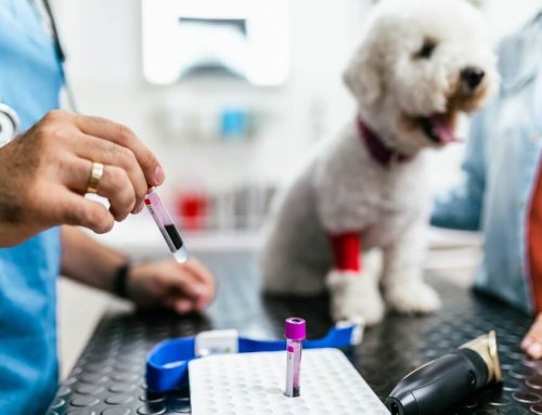
Our team at 360 Pet Medical offers several of the most commonly used advanced imaging modalities to diagnose your pet’s condition. We would like to provide information about these techniques and how we use their findings to make a diagnosis and develop a treatment plan for your pet.
Digital radiology in pets
An X-ray (i.e., radiograph) is a two-dimensional black-and-white image of an internal body area that is created when radiation passes through the area and captures the image. For a traditional radiograph, X-ray film generates the image by sensing the amount of radiation passing through the area and reaching the film. Dense tissue (e.g., bone) appears white on X-ray film, and less dense structures (e.g., lung tissue) appears black. Digital radiology is a more recent and advanced X-ray technology that captures images on a digital recording device and displays them on a computer screen. Digital X-ray images are typically higher in resolution than traditional X-rays, and the image can be manipulated to better view some body areas. In addition, your pet receives 80% less radiation through digital X-rays than traditional methods, making the digital technique safer for your pet. X-rays cause your pet no pain, but sedation may be required to prevent them from moving during the procedure, and ensuring a clear captured image. Digital X-rays commonly detect the following:
- Bone abnormalities — Fractured bones, bone loss in cases of infection, metabolic disorders, and bone cancer
- Joint abnormalities — Dislocation, and arthritis
- Foreign bodies — Foreign body ingestion
- Abnormal organs — Abnormal size, and the presence of lesions, indicating diseases such as cancer or pneumonia
- Internal bleeding — Often seen in trauma victims
Ultrasonography for pets
With ultrasound imaging, a transducer (i.e., probe) transmits sound waves through the body area of interest until they detect a boundary between tissues (e.g., between tissue and fluid, or tissue and bone). At the boundary, the sound waves reflect back to the transducer, recording the sound waves’ travel speed, direction, and distance, and generating a two-dimensional image. Ultrasound causes your pet no pain, and can usually be performed on a conscious animal. Different types of ultrasonography can diagnose many conditions, including:
- Abdominal ultrasound — Blood or urine test abnormalities may signal the need for an abdominal ultrasound, which allows our veterinary professionals to diagnose serious conditions such as pancreatitis, kidney or liver diseases, and various cancers. In addition, an abdominal ultrasound can confirm whether your pet has ingested a foreign object, if the material is not dense enough to appear on an X-ray. Abdominal ultrasound can also detect internal bleeding caused by trauma.
- Musculoskeletal ultrasound — If your veterinarian has been unable to diagnose your pet’s lameness through X-rays, which do not show soft tissue (i.e, tendons, ligaments, joint capsules, articular cartilage) changes well, they will perform a musculoskeletal ultrasound. These complementary imaging modalities—X-ray and ultrasound—are frequently used in tandem to determine a cause of lameness.
- Reproduction ultrasound — Ultrasound can determine whether your pet is pregnant, and can also be used to monitor fetal viability and development.
- Ocular ultrasound — Eye conditions (e.g., cataracts, uveitis) make visualizing the back of the eye difficult. Ultrasound can image this area to determine whether the retina is intact, the lens location is correct, and the amount of inflammatory debris inside the eye is normal.
- Thyroid ultrasound — Ultrasonography can help diagnose thyroid diseases in pets.
- Ultrasound-guided collection — Your veterinarian may suggest ultrasonography to obtain tissue samples less invasively than through open surgery. Fine-needle lymph node or tumor aspiration is more successful when performed using ultrasonography, enabling biopsies without leaving a large surgical incision. Ultrasound-guided collection is typically performed while your pet is under general anesthesia or heavy sedation.
Echocardiography in pets

An echocardiogram is a specific ultrasound that provides information about the heart’s size, shape, and functional ability. Through this modality, a veterinary echocardiologist can observe the heart’s structures (i.e., the four chambers, heart wall, valves, pericardial sac). Color Doppler can assess how blood enters, exits, and flows through your pet’s heart. This procedure is technically difficult, requiring the use of a specialized cardiac transducer by a veterinary echocardiologist who has advanced training. We typically recommend an echocardiogram for pets who are at high risk of heart disease based on breed or size, have a heart murmur, a suspected heart condition, a persistent cough, or a fainting tendency. Dr. Loni Odenbeck is one of only a small handful of veterinarians specifically trained in performing echocardiograms in Southwest Montana. There is currently no board-certified cardiologist practicing in the state of Montana. Once the images are obtained, a telemedicine consultation with a board-certified cardiologist can be done to discuss treatment plans, further diagnostics or monitoring parameters.
Advanced imaging techniques can be extremely useful for diagnosing certain pet conditions. If your pet is showing disease signs, contact our team at 360 Pet Medical, so we can determine whether your pet should undergo an advanced imaging procedure.


Leave A Comment